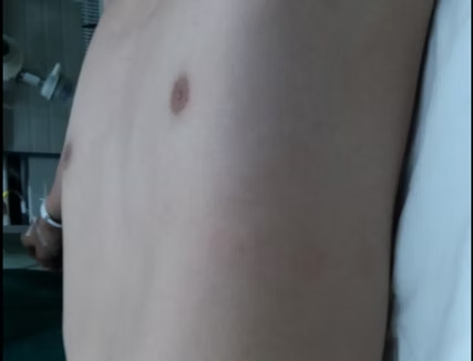
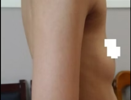
Flat chest is a common chest wall deformity characterized by a flattened appearance of the chest wall, with a significantly shortened anteroposterior diameter that results in a measurement less than half of the transverse diameter.
Introduction
Causes
The exact cause of flat chest is not clear yet. It may be related to genetic factors or malnutrition, and can also be caused by chest trauma, chronic wasting diseases, or pulmonary diseases.
Symptoms
Flat chest can compress the heart and lungs, potentially causing symptoms such as palpitations, shortness of breath, and chest tightness. This condition can also lead to significant psychological stress, often making patients feel quite inferior and potentially resulting in severe psychological issues.
Furthermore, patients with flat chest frequently experience spontaneous pneumothorax.
Diagnostic Methods
Diagnosis primarily involves a physical examination by observing the appearance of the chest wall, supplemented by imaging examination such as X-ray and chest CT scan.
Surgical Procedures
This new surgical method, introduced as an improvement over the Nuss procedure, retains the basic operating principle but integrates innovative and optimized methods at each stage. The Wung procedure employs many unique surgical techniques, including safer and more straightforward methods for the placement and fixation of the bar. These innovations not only greatly enhance the safety, reliability, and effectiveness of the surgery, but also reduce the incidence of complications and shorten the patient’s postoperative recovery time.
Frequently Asked Questions
Flat chest is characterized by a significantly reduced anteroposterior diameter of the thorax, resulting in a flattened chest wall with loss of natural curvature. In contrast, individuals with a slender build have a normal anteroposterior chest diameter and maintain the natural contour and curvature of the chest wall.
No, flat chest does not improve spontaneously and may worsen with age. Compared to pectus excavatum, flat chest often involves a broader area of depression, which can impose more severe compression on the heart and lungs. If patients experience symptoms like palpitations, shortness of breath, or chest tightness, surgical intervention is generally recommended.
Typically, 2 small incisions are made, one on each side of the chest.
In most cases, 2 to 3 bars are required. The exact number depends on the patient’s condition and the surgical plan.
In most cases, the deformity is significantly corrected right after the procedure, resulting in a chest wall that appears close to normal. Following this initial correction, the chest continues a process of slight, gradual remodeling over time. The new contour is typically well-established within about 3 months and fully stabilizes after 2 to 3 years.
The total cost usually ranges from $7,000 to $14,000. The exact amount will be determined by factors such as the patient’s condition and the specific surgical plan.
It is common to experience significant pain in the initial postoperative period, particularly among adolescent and adult patients due to their more rigid skeletal structure. Our hospital employs a comprehensive, multi-modal analgesia protocol to ensure effective pain control. This integrated approach includes:
- Intraoperative Intervention: Intercostal nerve blocks are administered to block pain transmission.
- Postoperative Medication: Continuous pain management is delivered through a patient-controlled analgesia (PCA) pump, supplemented with scheduled intravenous analgesics.
- Adjunctive Rehabilitation Therapy: Our dedicated rehabilitation team provides personalized physiotherapy, incorporating techniques such as acupuncture, therapeutic massage, electrical stimulation, and ultrasound therapy. These modalities are highly effective in alleviating localized pain and common discomforts like postoperative bloating.
Most patients stay in the hospital for around 7-10 days, although the actual duration depends on individual recovery.
The risk is very low. The bars used in surgery are made of titanium alloy, which provides excellent rigidity and resistance to deformation. In addition, the Wang Technique, a cutting-edge bar fixation method, is utilized during surgery to rigidly stabilize the bars in position, effectively preventing displacement. Long-term clinical data confirms that the vast majority of patients do not experience bar displacement or deformation. It is crucial to note that during the early postoperative period (within the first 3 months), patients should avoid vigorous exercise and be mindful in their daily lives to avoid significant impact or trauma to the chest, thereby reducing the likelihood of bar displacement.
Most patients can get out of bed and walk within 4–5 days after surgery, and resume daily activities around 10 days postoperatively. Patients can usually return to normal work or school (excluding heavy physical labor) around 1 month. Light exercise, such as jogging or hiking, can start within the first three months, with intensity gradually increased thereafter.
It is crucial to note that if you encounter any discomfort, such as chest pain or shortness of breath, during exercise, you should stop the activity immediately. If necessary, a chest X - ray or CT scan can be arranged for further examination.
Yes, for the initial postoperative period, maintaining a specific sleeping position is important for healing. It is advised to sleep in a supine position (lying on your back) or a modified lateral position (partially reclining on one side) for the first month , adjusting as needed for comfort around the incision sites. Additionally, you need to avoid movements with a large range of motion, like chest expansion, bending over, and lifting heavy objects. After about a month, once your incisions have completely healed, you can gradually start sleeping on your side.
After the drainage tubes are removed (the removal time is determined by the drainage volume and follow - up examination results, generally within 1 - 2 weeks), you may shower with waterproof dressings covering the incision, then replace them with breathable gauze afterwards. Around 3 weeks after surgery, once the incisions have completely healed, you can shower normally.
Our discharge protocol is designed to ensure your safety. Typically, patients can be safely discharged 10 - 14 days post-surgery. After about one week of observation post-discharge, the risk of the vast majority of complications can generally be excluded. If recovery progresses smoothly during the first three weeks post-surgery, the likelihood of later complications is extremely low. Additionally, the bars are firmly secured, and the surgical technique is specifically designed to prevent issues like bar displacement.
However, if you notice symptoms such as pneumothorax, pleural effusion, significant pain, or poor wound healing, please contact our doctors promptly. In case of an emergency, please seek immediate care at a local hospital.
If your recovery goes well without any noticeable discomfort or abnormal conditions, regular follow-up is usually not required. However, if you develop symptoms such as persistent high fever (temperature >38.5°C), sudden chest tightness, shortness of breath, or difficulty breathing, please have a chest X-ray or CT scan locally and consult a thoracic surgeon or our doctor for further guidance.
The bars are usually removed three years after surgery, with the exact timing determined by the patient’s recovery and the doctor’s evaluation.
Yes. Upon discharge, we will provide a discharge summary and a medical certificate. If airport security raises any concerns, presenting these medical documents will facilitate your passage.
In most cases, taking a flight does not cause discomfort, you can travel with confidence.
Optimal nutrition is crucial for healing. We recommend the following dietary plan, tailored to your recovery phase:
Initial Phase (during bed rest): Choose easily digestible semi-liquid foods, such as porridge, juice, or well-cooked noodles.
Recovery Phase (once able to get out of bed): Gradually resume a normal, balanced diet with an emphasis on high-quality protein sources such as fish, chicken, and eggs. Make sure to eat fresh fruits and vegetables daily to supplement vitamins and electrolytes.
Precautions: Strictly avoid spicy and greasy foods, and be cautious with foods that may trigger allergies, such as seafood or mangoes.